Benign Does Not Mean Simple: From CT Imaging to Complex Wound Closure at Pet ER
Advanced imaging, mass excision, negative-pressure wound therapy, ICU monitoring, and hyperbaric oxygen therapy—all coordinated under one roof.
Benign Does Not Mean Simple: From CT Imaging to Complex Wound Closure at Pet ER
A large mass may appear straightforward from the outside. Surgically, however, its true size, depth, relationship to surrounding structures, and effect on the available skin may make treatment considerably more complex.
This senior canine patient presented to Pet ER Small Animal Emergency Hospital with extensive swelling of the right hind limb and an open wound over a long-standing mass. Because the abnormality involved a large portion of the limb, detailed diagnostic imaging was essential before attempting surgery.
Imaging Before Incision
At Pet ER, complex surgery begins with careful diagnostic planning.
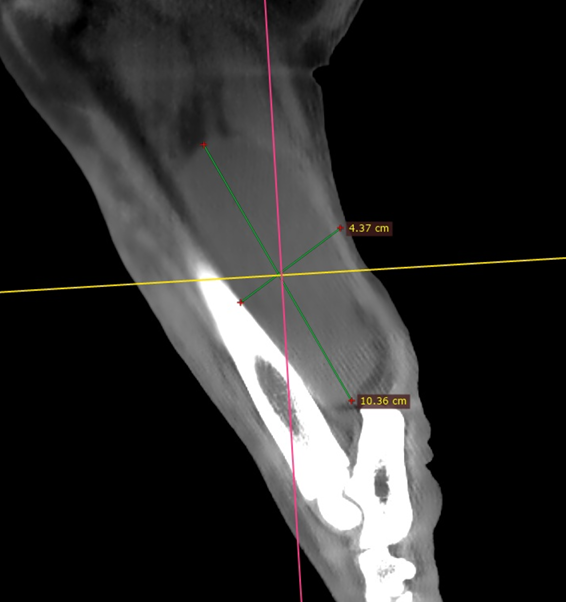
CT imaging was performed to define the size and location of the abnormalities and evaluate their relationship to the muscles, bones, joints, and calcaneal tendon.
The CT examination identified two significant findings within the right hind limb:
• A large fat-attenuating mass within the caudomedial thigh muscles, measuring approximately 11 cm in height and 3.7 cm in width.
• A separate soft-tissue-to-fluid-attenuating mass near the distal tibia, measuring approximately 10 cm by 4.4 cm and displacing the calcaneal tendon.
The imaging also documented severe degenerative disease affecting both stifle joints.
This information allowed the surgical team to understand that the visible swelling was not simply a superficial lump. Advanced imaging helped establish the extent of the disease, guide incision planning, anticipate the tissue deficit that could remain after excision, and discuss the expected recovery with the patient’s family.
🩺 Complex Surgical Excision
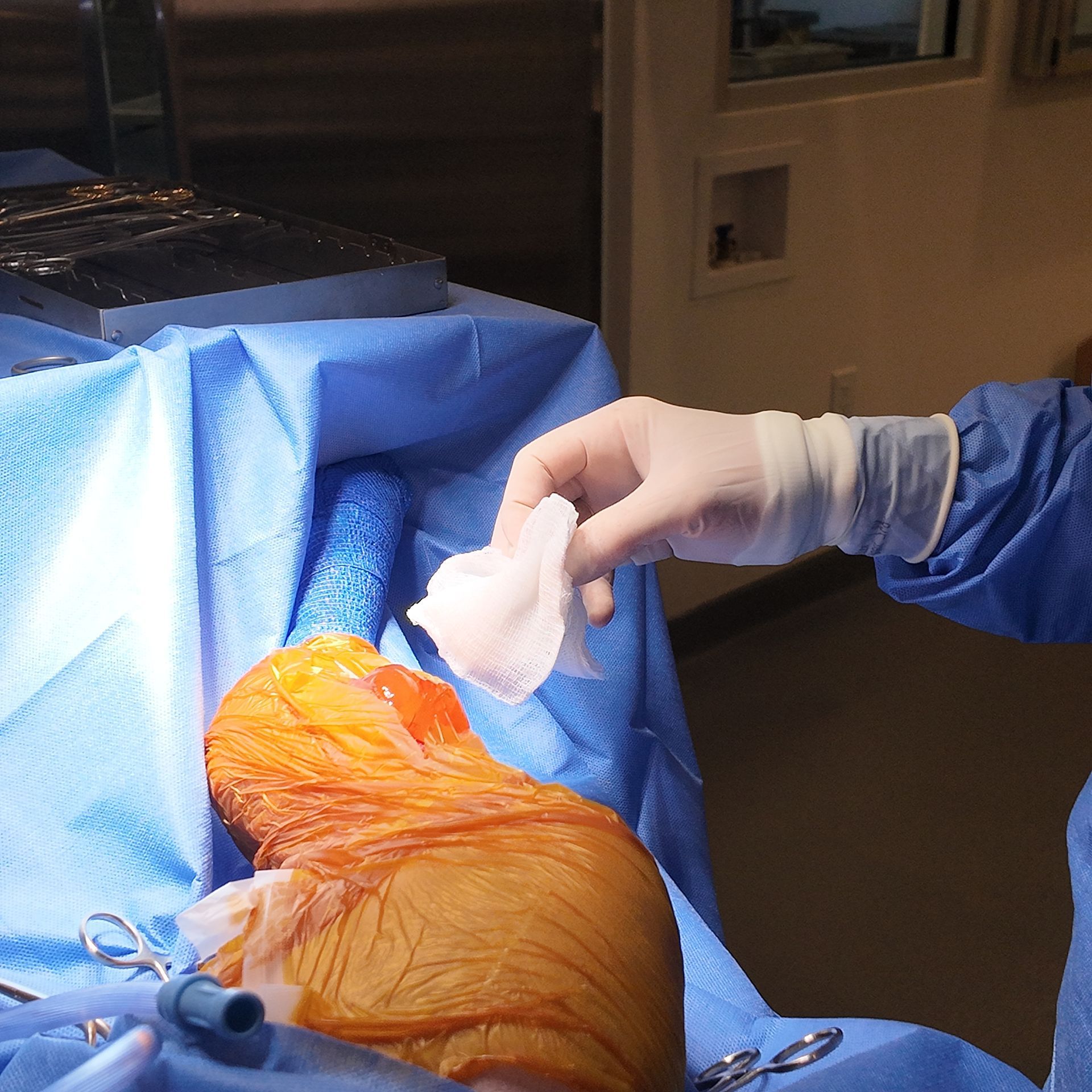
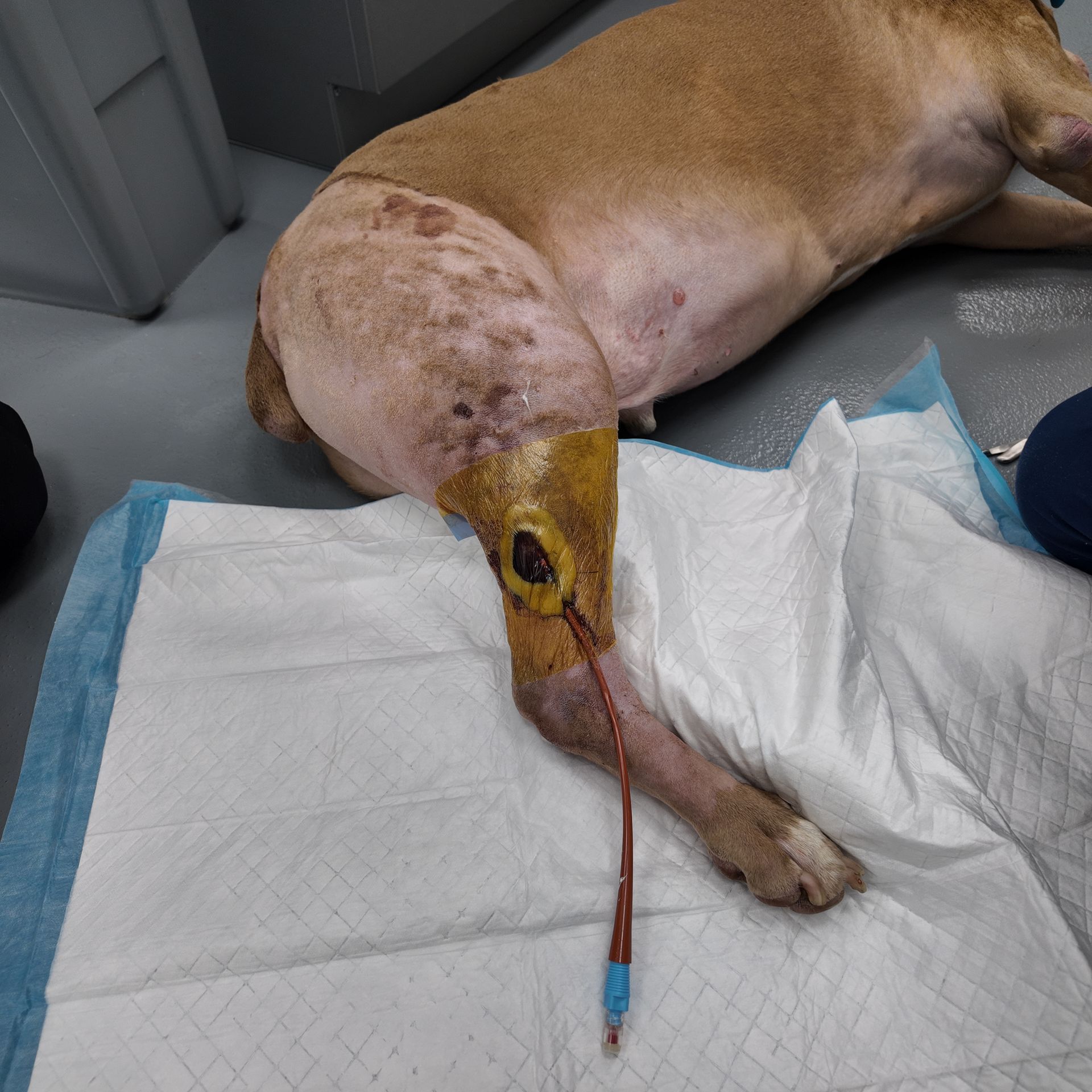
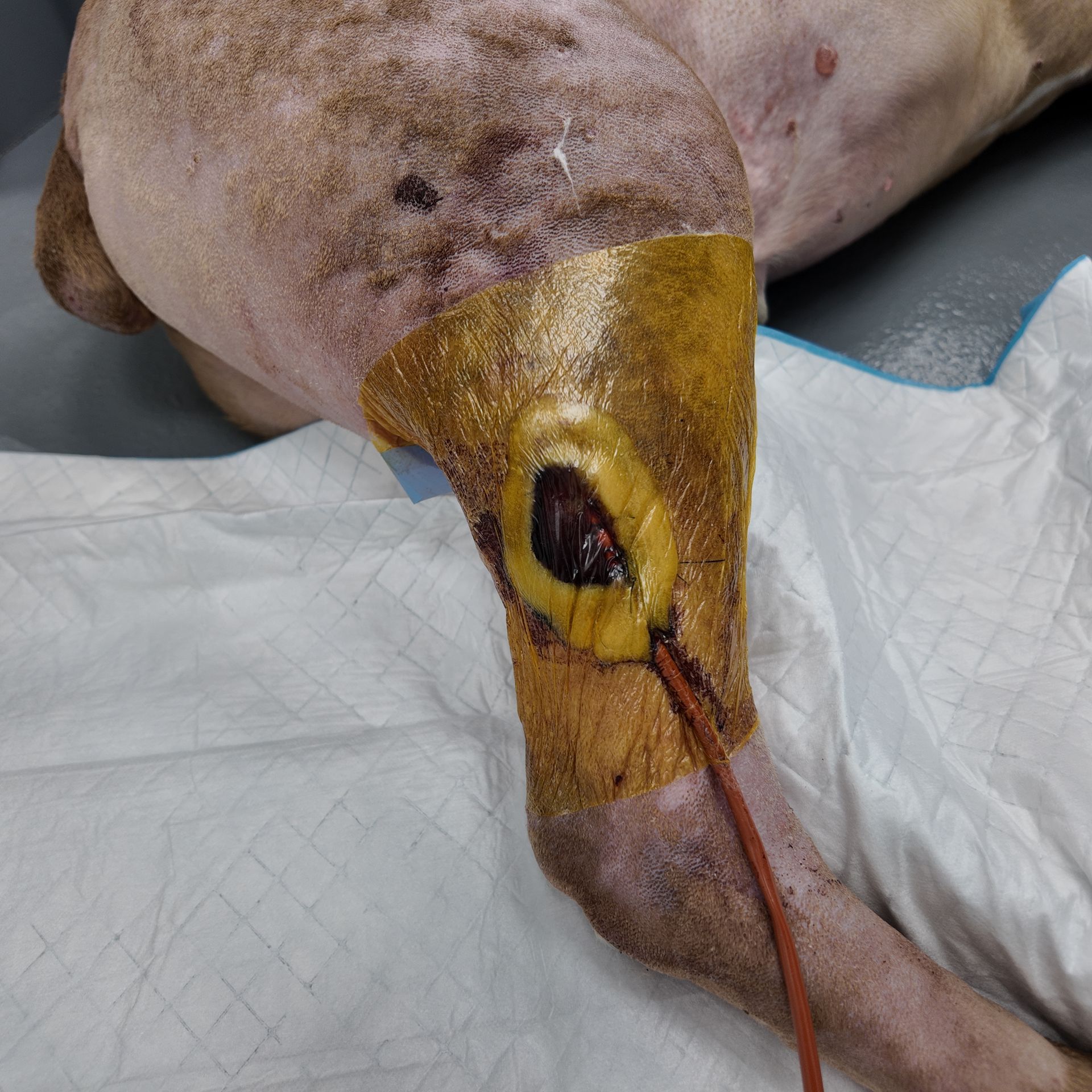
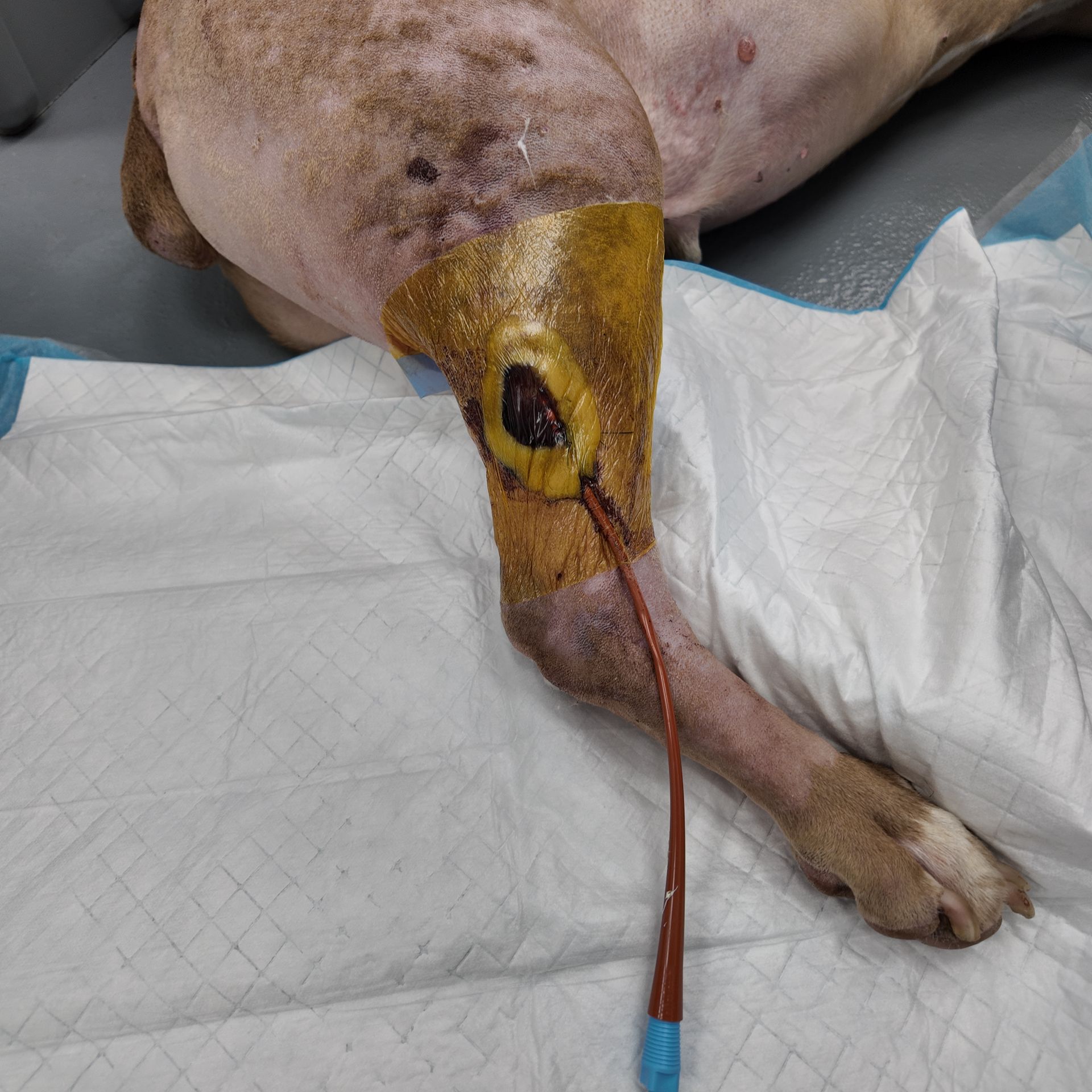
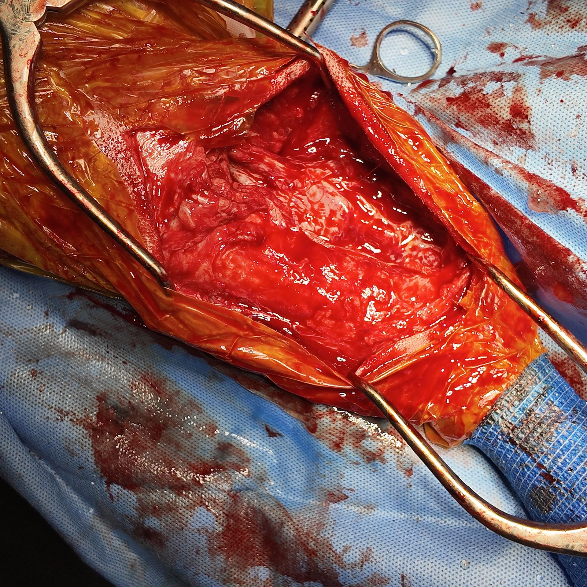
The abnormal tissue was surgically exposed and excised.
With a lesion of this size, successful surgery is not limited to removing the mass. The surgeon must also protect important surrounding structures, manage bleeding, preserve viable tissue, and determine how the remaining wound can be closed safely.
After excision, a substantial tissue void remained. There was also insufficient healthy skin available to bring the wound edges together with a routine tension-free closure.
Attempting to force an immediate closure in that situation could compromise the remaining tissue. The wound therefore required a staged and more advanced management plan.
Vacuum-Assisted Wound Closure
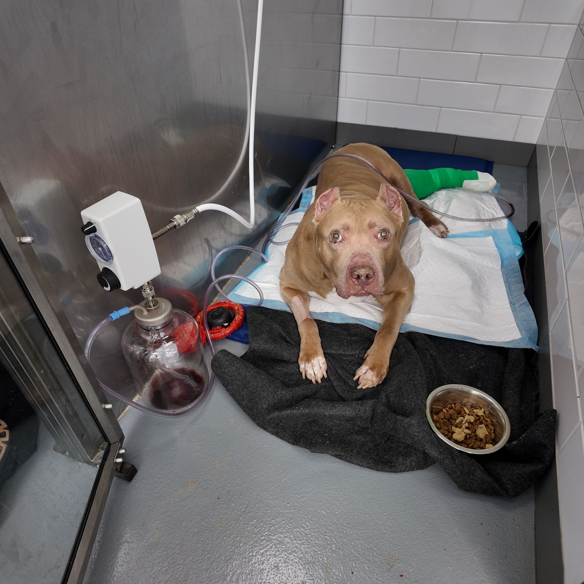
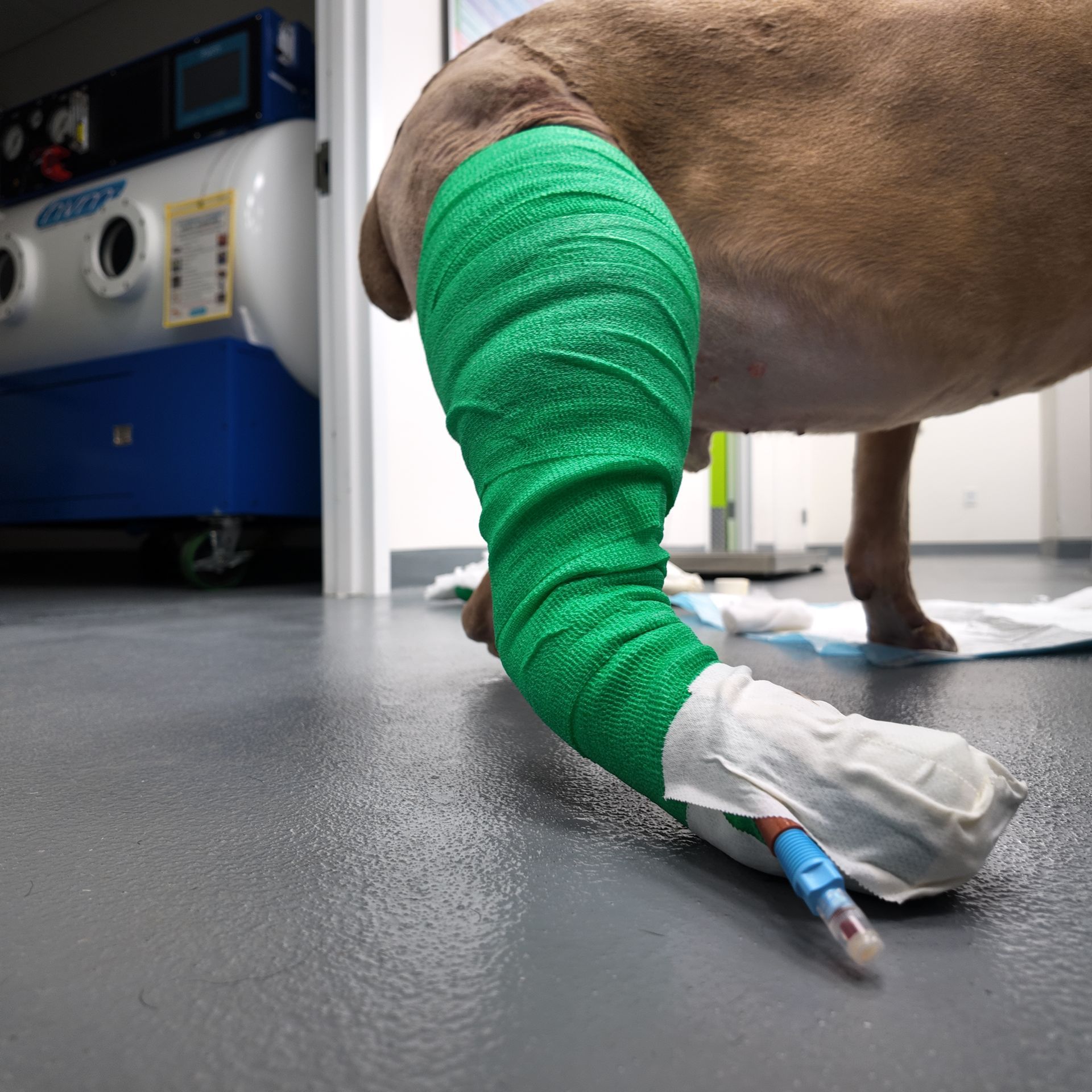
Negative-pressure wound therapy, also known as vacuum-assisted wound closure or wound VAC therapy, was used to manage the postoperative wound.
This system applies controlled negative pressure to the wound through a specialized dressing. In this case, it allowed the Pet ER team to manage wound fluid, protect the surgical bed, reduce the effects of the large remaining tissue space, and support the wound as it progressed toward closure.
The ability to provide negative-pressure wound therapy in-house was especially important because the treatment required continued device management, repeated evaluation, bandage care, and close monitoring.
ICU Hospitalization and Continuous Wound Management
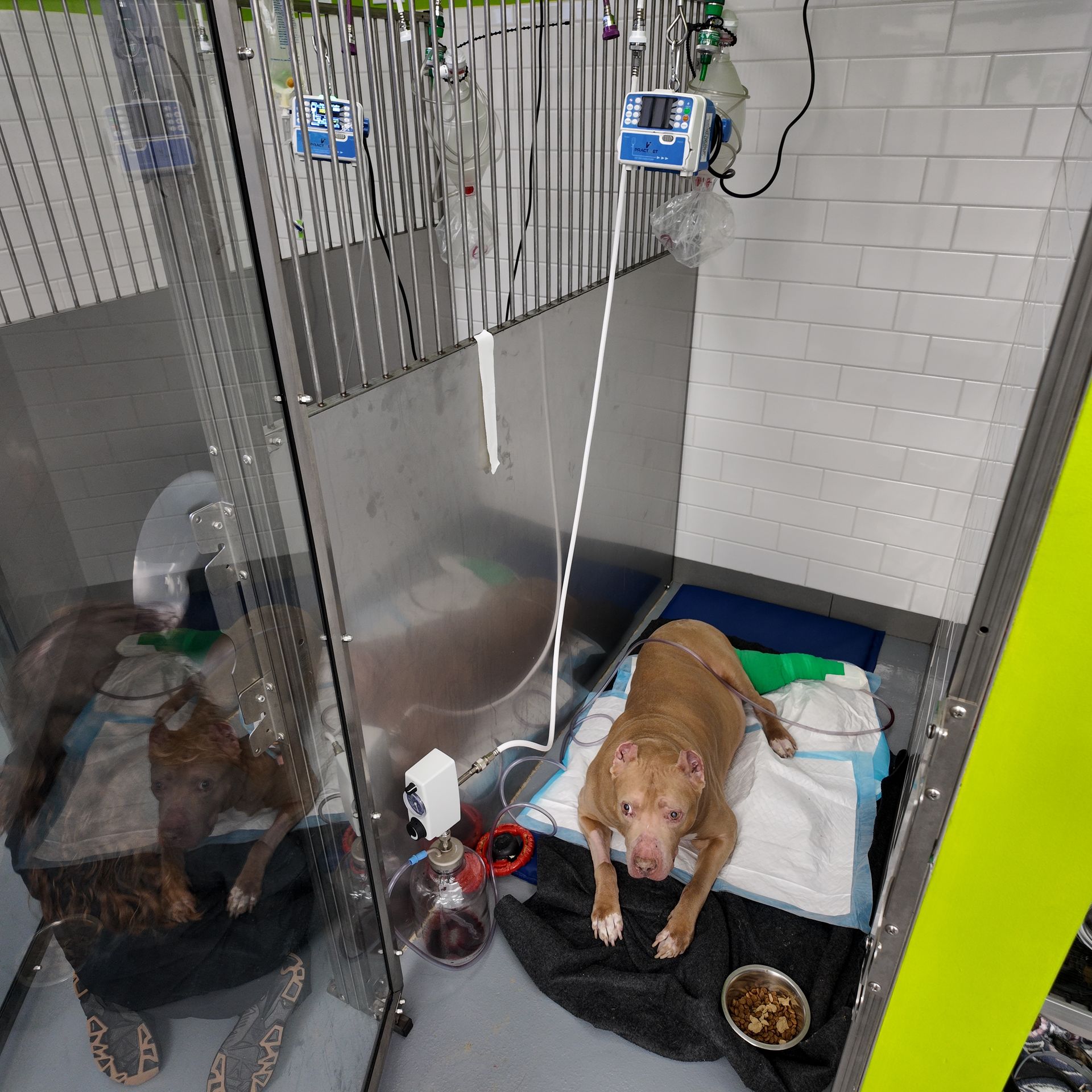
The patient remained hospitalized in the Pet ER ICU during this phase of recovery.
ICU care allowed our team to provide:
• Continuous patient monitoring
• Pain management
• Repeated assessment of the surgical site
• Vacuum-assisted wound therapy management
• Bandage and drain care
• Nutritional and mobility support
• Immediate intervention if the wound or the patient’s condition changed
Complex wound cases do not end when the patient leaves the operating room. The postoperative period is an active continuation of the surgical plan, and careful ICU management can be just as important as the operation itself.
Hyperbaric Oxygen Therapy
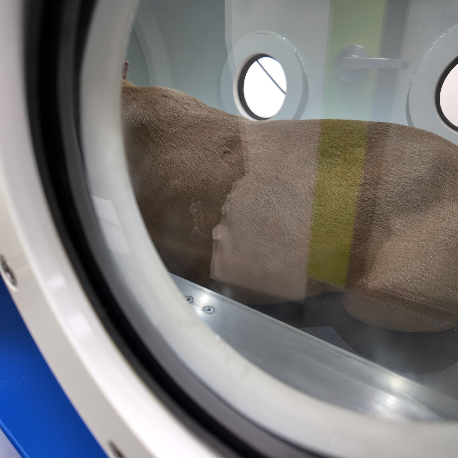
Hyperbaric oxygen therapy was also incorporated as an adjunct to the patient’s recovery plan.
During hyperbaric treatment, the patient breathes oxygen within a pressurized chamber. For selected patients, this can be used as part of a broader wound-care strategy to support oxygen delivery to healing tissues.
Because Pet ER maintains its own veterinary hyperbaric oxygen chamber, this therapy could be coordinated with the patient’s surgery, ICU hospitalization, negative-pressure wound therapy, and serial wound assessments without transferring the patient between multiple facilities.
The Final Diagnosis
Histopathology ultimately confirmed that the excised cystic lesion was a benign hygroma, with no evidence of neoplasia.
A hygroma is an acquired, fluid-containing structure that can develop over an area exposed to repeated pressure or trauma. Although the diagnosis was benign, the lesion’s size, location, chronicity, effect on surrounding tissues, and resulting skin deficit made the case anything but minor.
This case is an important reminder that “benign” does not necessarily mean “simple.”
A noncancerous lesion may still require advanced imaging, extensive surgery, complex wound reconstruction, ICU hospitalization, and prolonged postoperative management.
🐾 One Coordinated Continuum of Care
This patient’s treatment incorporated:
CT imaging
Complex surgical excision
Negative-pressure wound therapy
ICU hospitalization
Hyperbaric oxygen therapy
Histopathology
All stages were coordinated at Pet ER Small Animal Emergency Hospital.
Advanced veterinary care is not defined by owning one piece of equipment or performing one procedure. It is the ability to connect diagnostics, surgery, wound management, critical care, and recovery into one continuous medical plan.
That is the level of integrated care Pet ER was built to provide.
Every patient and every condition is different. Treatment recommendations depend on the patient’s examination, diagnostic findings, overall health, and individual surgical needs.
For emergency surgery, advanced imaging, complex wound management, or ICU care, please call Pet ER at (631) 247-3837 to confirm current doctor availability and emergency intake.