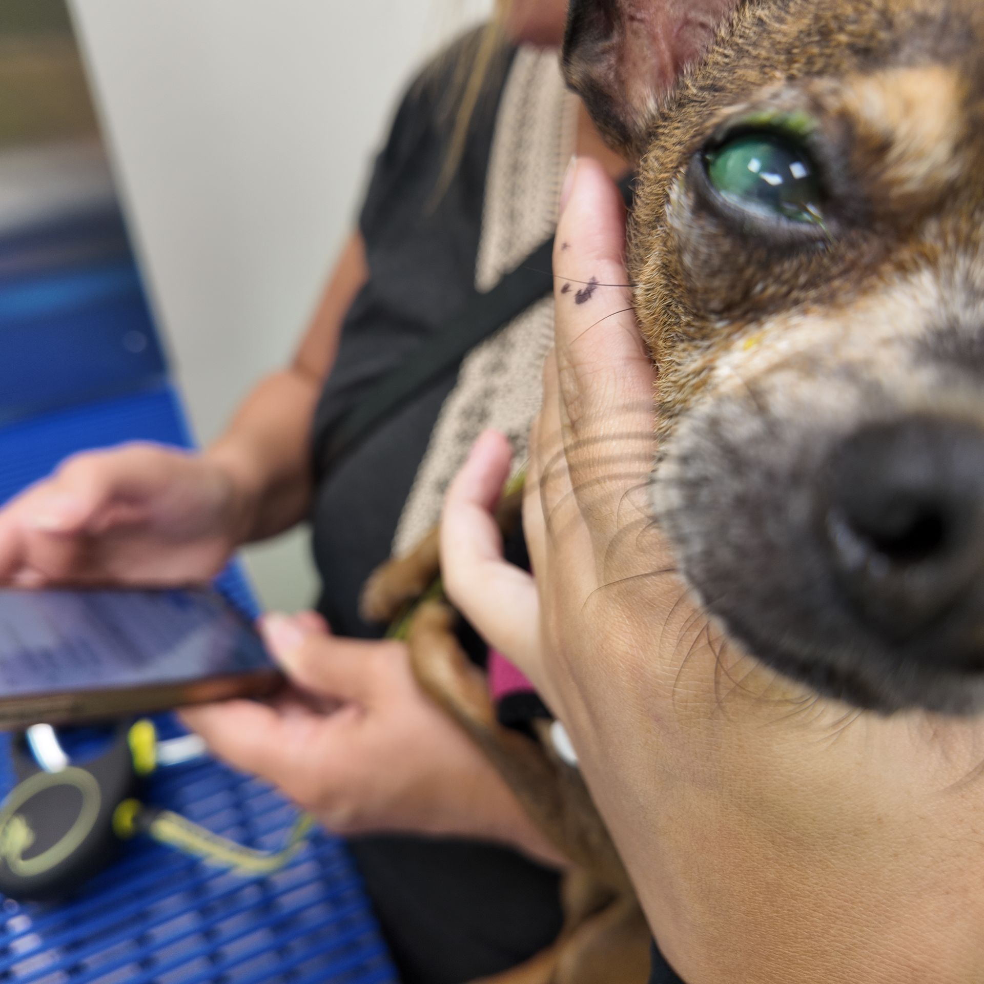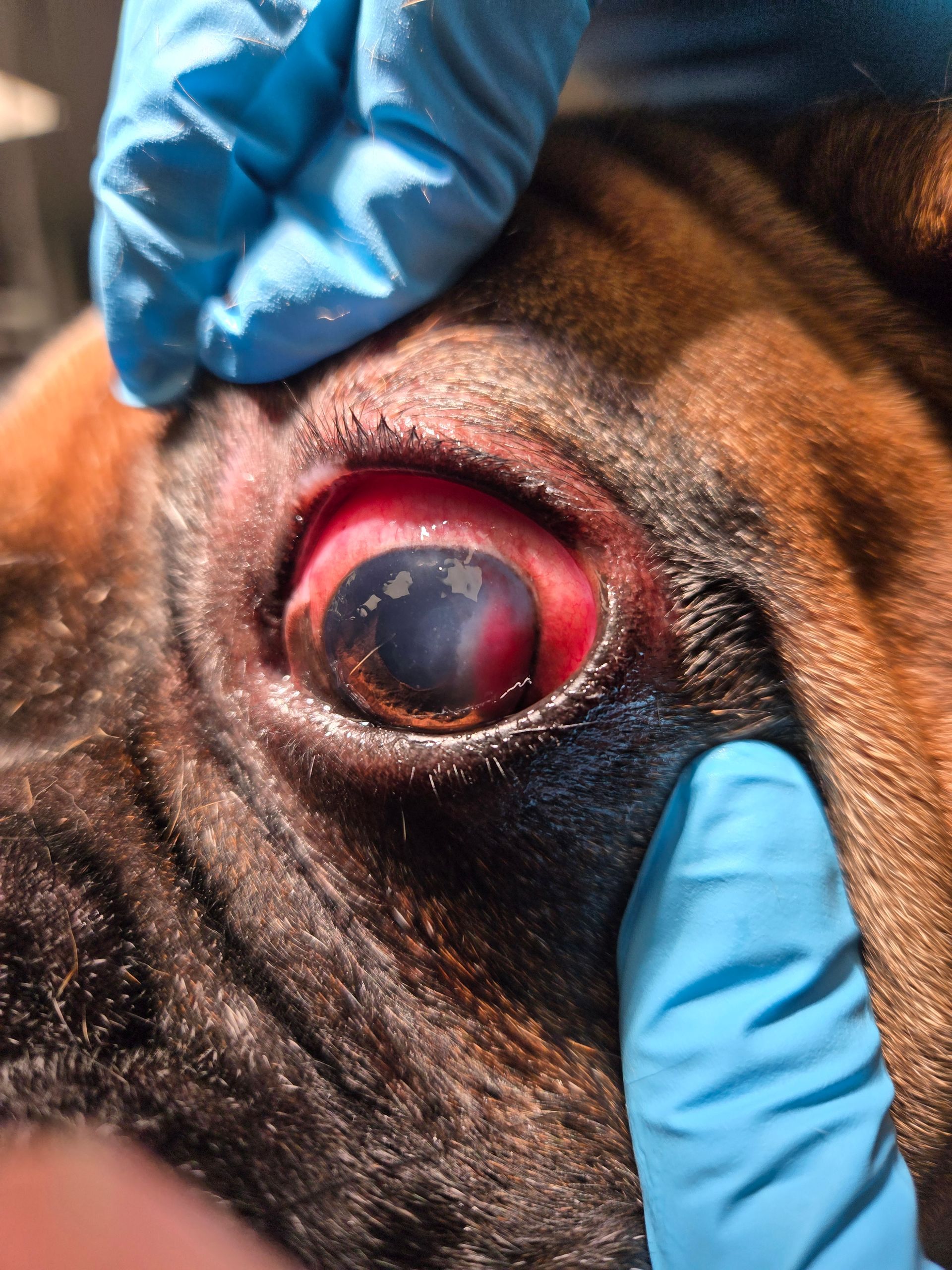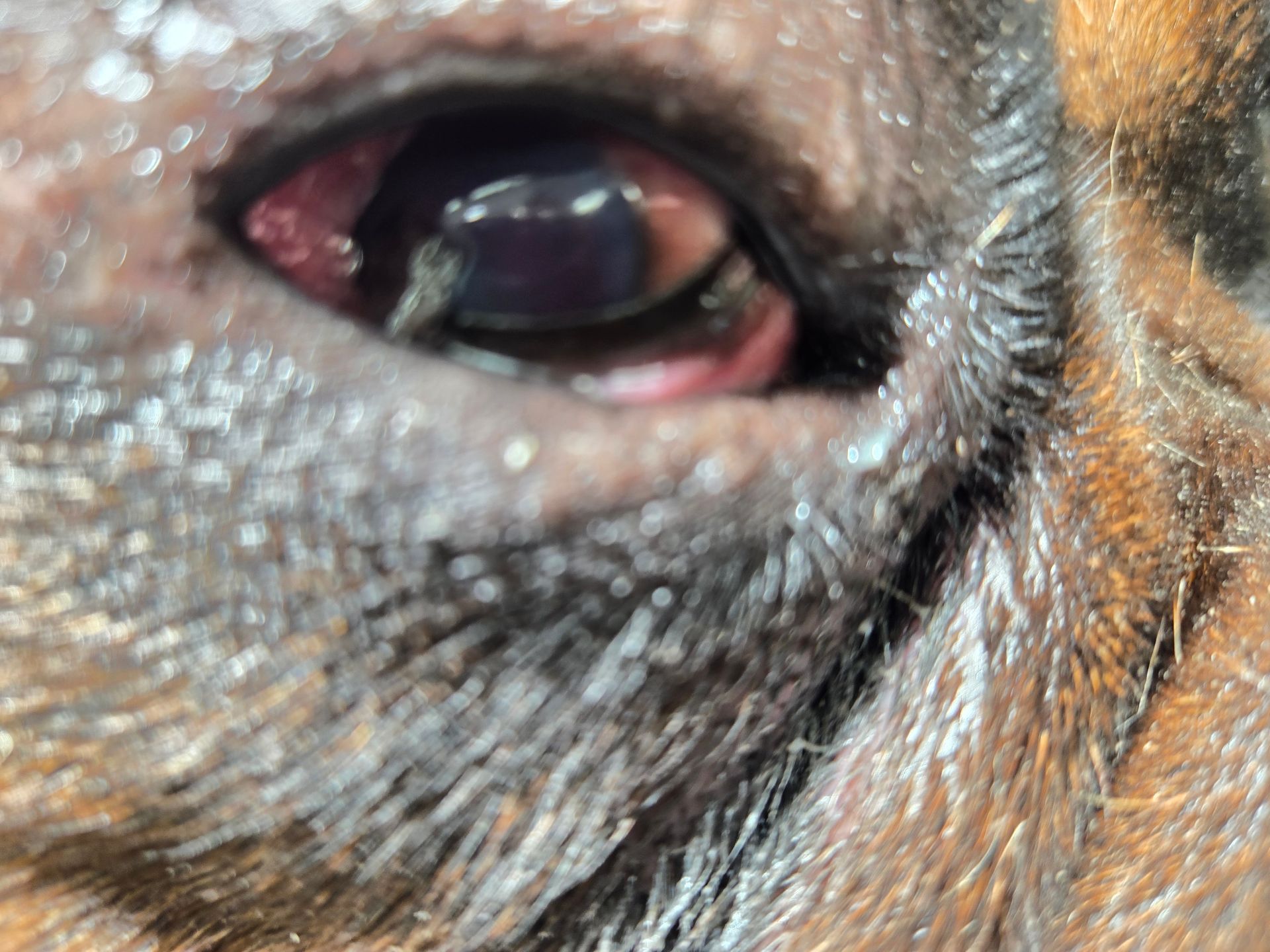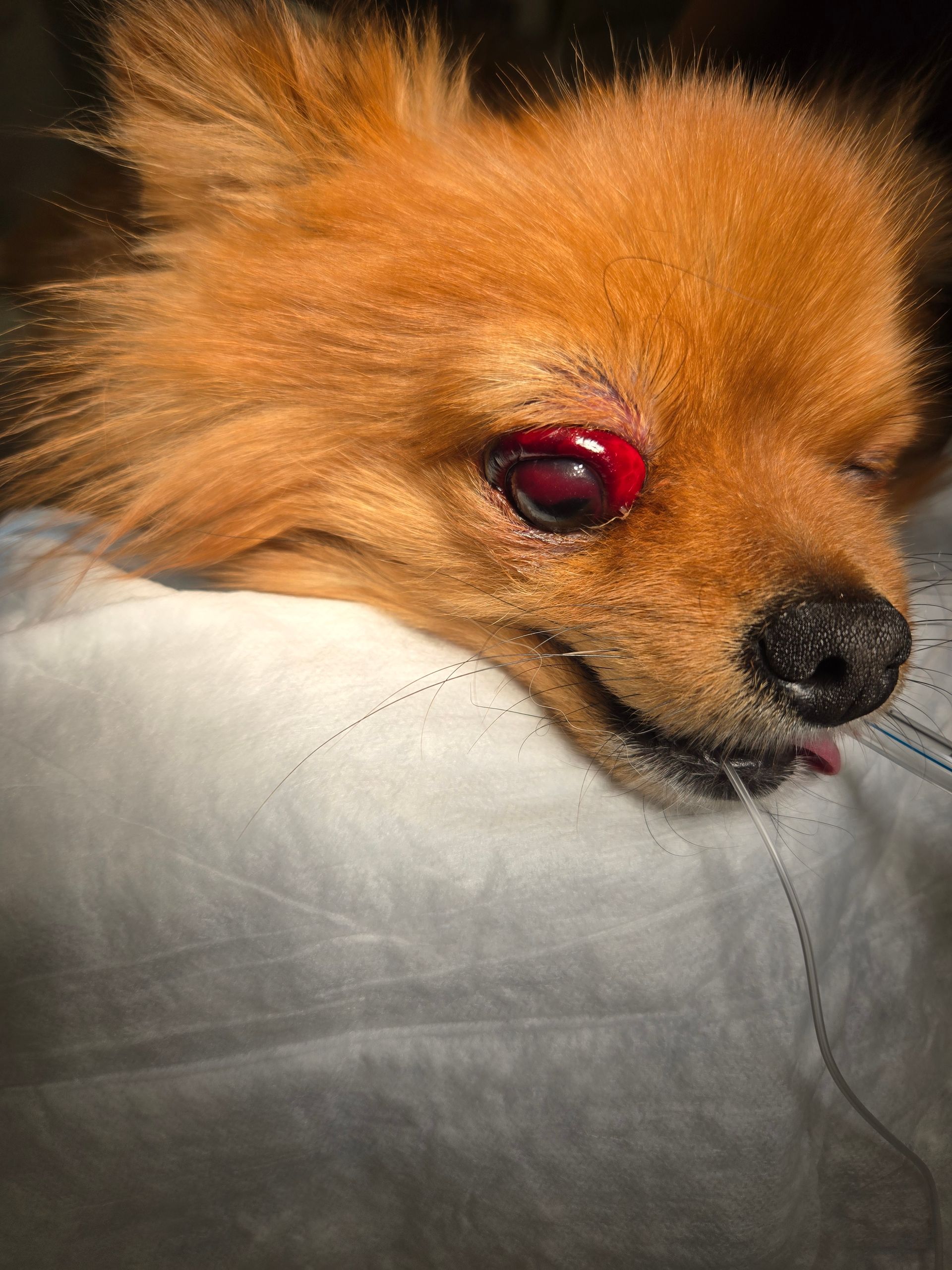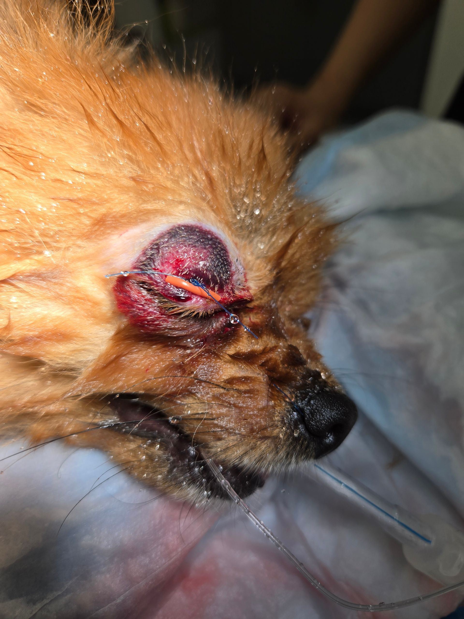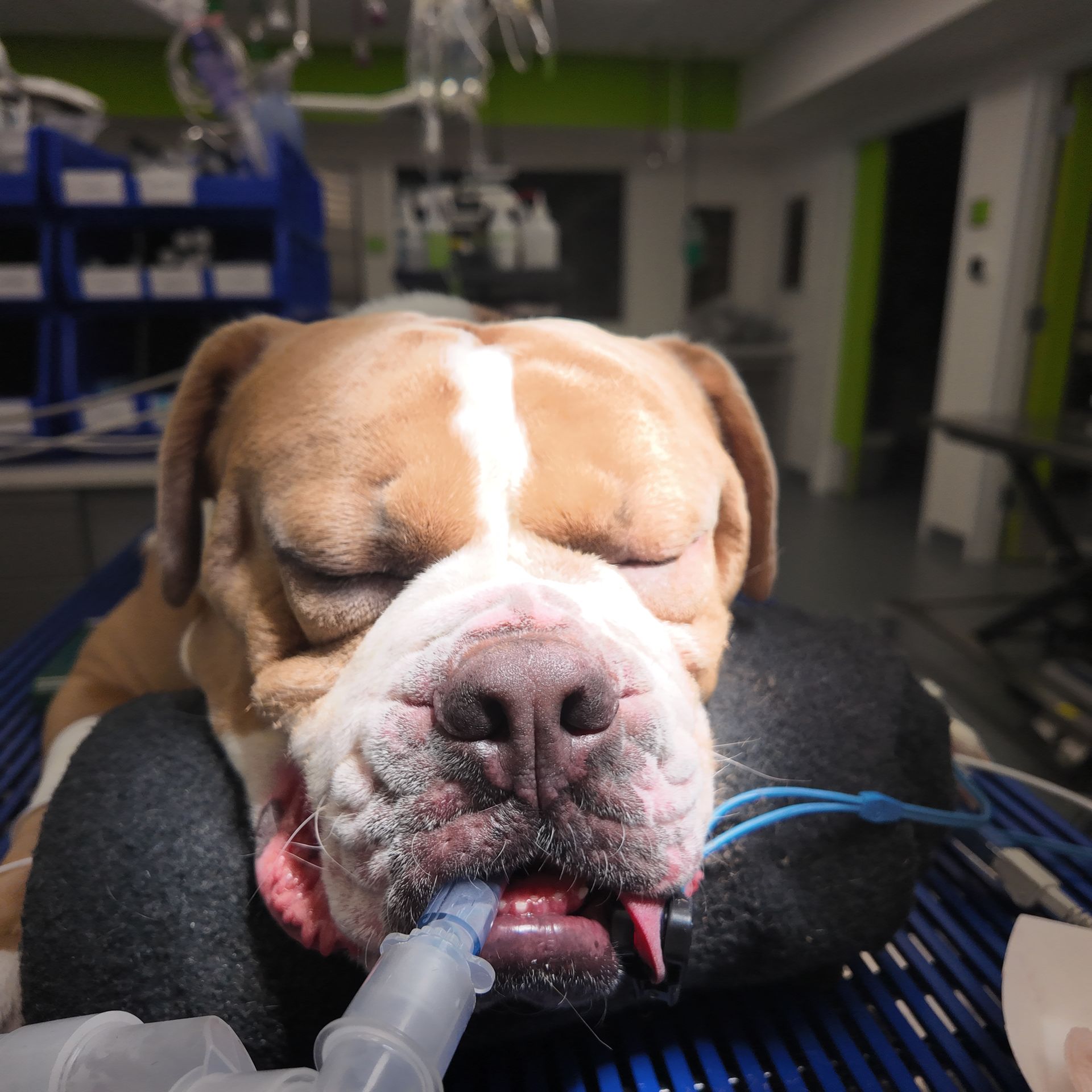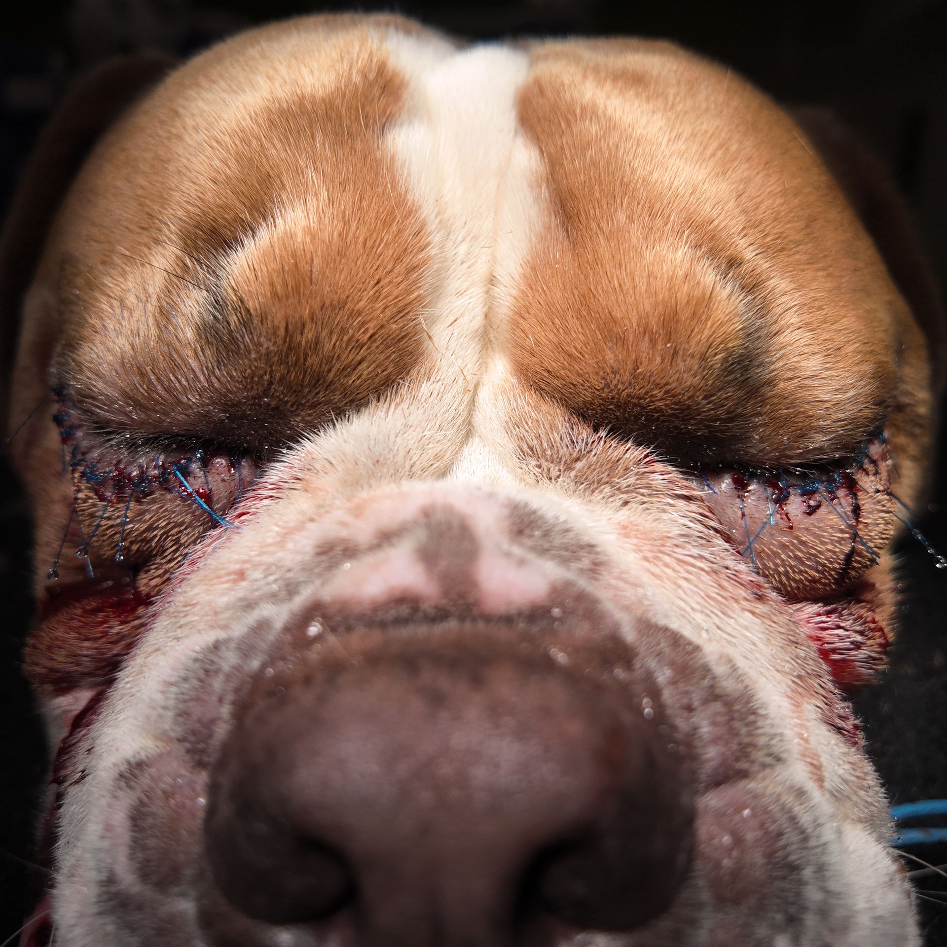
👁️ Real emergency patients. Real vision-threatening conditions. Real emergency care. 🐾
From severe ocular trauma to surgical eye protection, Pet ER manages time-sensitive eye emergencies with urgency and precision.
WHEN MINUTES MATTER: EMERGENCY EYE CARE AND OCULAR ULTRASOUND AT PET ER
Serious ocular trauma, emergency eye procedures, advanced imaging, and follow-up care for dogs and cats.
⚠️ This article contains clinical photographs of serious eye injuries and emergency treatment.
Eye emergencies can deteriorate rapidly. A dog or cat with a suddenly red, swollen, protruding, cloudy, painful, bleeding, or injured eye should not be treated as a routine “wait and see” case.
At Pet ER Small Animal Emergency Hospital, we provide urgent evaluation, medical stabilization, ocular ultrasound, and selected emergency eye procedures for dogs and cats throughout East Setauket, Suffolk County, and Long Island.
Our goal is to act quickly, relieve pain, protect delicate ocular tissues, determine the extent of the injury, and give each patient the best possible opportunity for comfort, healing, and preservation of the eye whenever medically appropriate.
WHY OCULAR EMERGENCIES CANNOT WAIT
The eye contains delicate structures that may be permanently damaged by trauma, pressure, exposure, inflammation, infection, or loss of normal blood supply.
A pet may otherwise appear stable while the eye itself is experiencing a true emergency. Delaying evaluation can increase pain, worsen tissue injury, reduce the possibility of preserving vision, and in severe cases place the entire eye at risk.
Emergency warning signs may include:
• A protruding or displaced eye
• Sudden severe redness
• Marked swelling around the eye
• Squinting or holding the eye closed
• Cloudiness or a blue-gray appearance
• Blood inside or around the eye
• Visible injury to the eyelid or surrounding tissues
• Sudden vision loss or disorientation
• Unequal pupils
• Discharge associated with pain
• Trauma from another animal, a vehicle, a fall, or a foreign object
• Pawing or rubbing at the face
• A rapid change in the position, shape, or appearance of the eye
A painful or suddenly abnormal eye should be considered an emergency until proven otherwise.
EMERGENCY OCULAR ASSESSMENT
Emergency eye care begins with a rapid assessment of the patient as a whole. Trauma involving the eye may also involve the head, airway, brain, chest, or other areas of the body, so stabilization of life-threatening problems always comes first.
Once the patient is stable, our team carefully evaluates the eye, eyelids, surrounding tissues, and visual responses. Depending on the condition and the patient’s tolerance, the evaluation may include:
• Examination of the globe and eyelids
• Assessment of pupil responses and vision
• Fluorescein staining to identify corneal injury
• Measurement of intraocular pressure when appropriate
• Assessment for bleeding, exposure, swelling, and tissue viability
• Evaluation of the surrounding bones and soft tissues
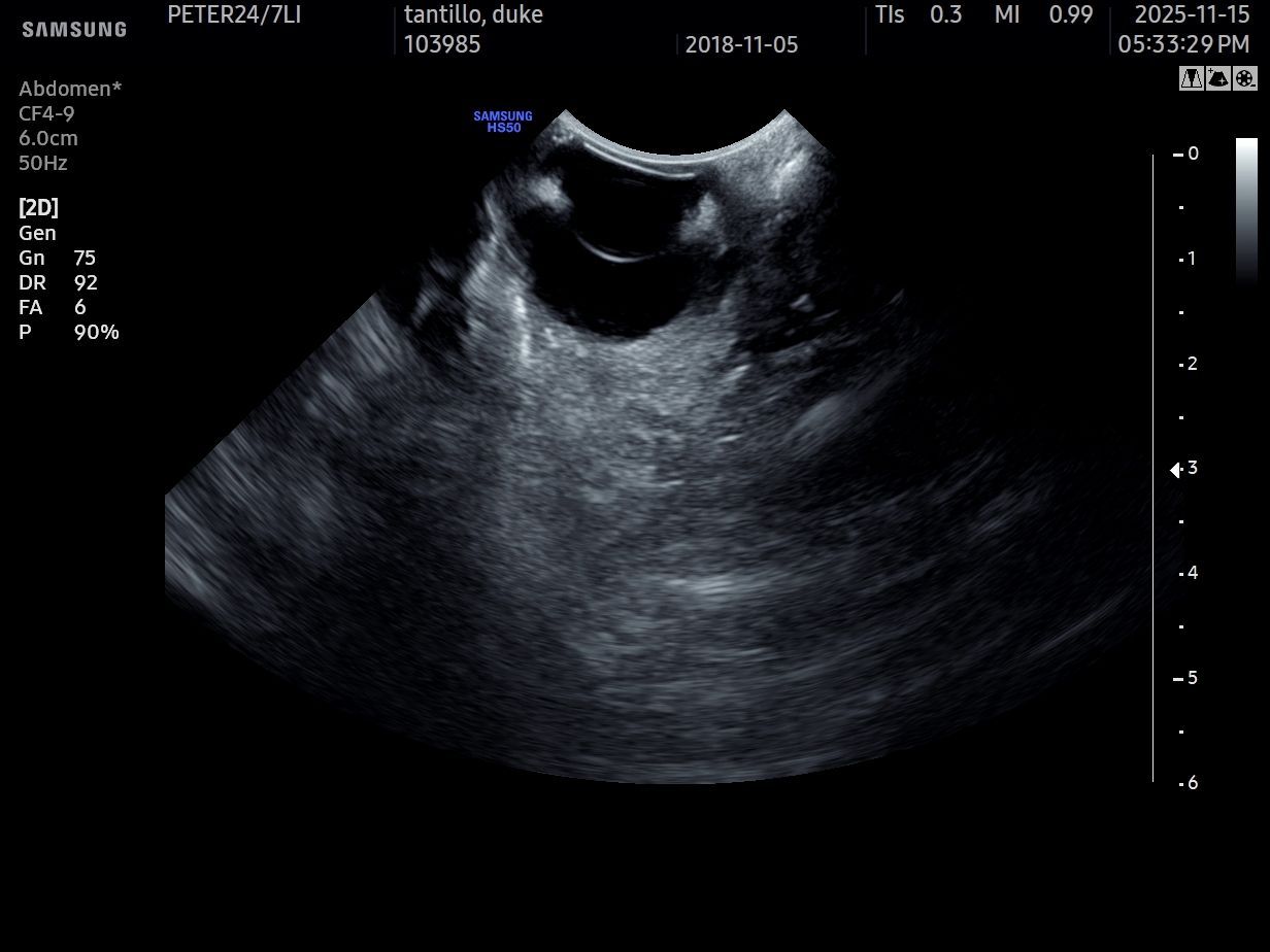
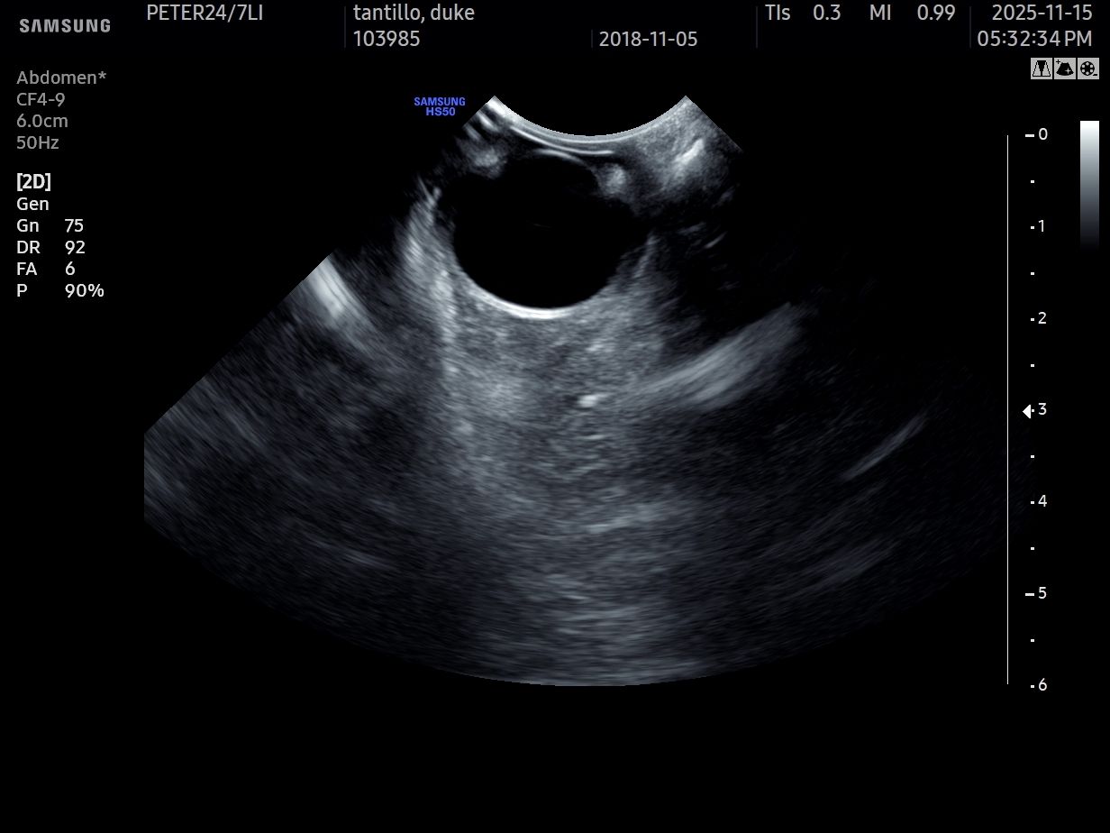
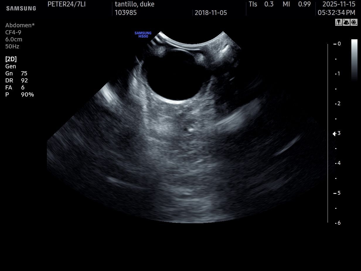
• Ocular ultrasound when the internal eye cannot be fully visualized
Eye injuries are often extremely painful. Sedation or anesthesia may be necessary to allow a complete examination and safe treatment without causing additional distress or damage.
OCULAR ULTRASOUND: SEEING BEYOND A CLOUDY OR INJURED EYE
When we cannot see clearly into the eye, we do not simply guess—we use diagnostic imaging when clinically appropriate.
Blood within the eye, severe corneal swelling, cloudiness, cataract, inflammation, trauma, or distortion of the globe may prevent direct visualization of the internal ocular structures. In these situations, ocular ultrasound can provide valuable information that cannot be obtained from the external examination alone.
When safe to perform, ocular ultrasonography may help evaluate:
• The lens and its position
• The vitreous chamber
• The retina
• The contour and integrity of the globe
• Internal hemorrhage or abnormal material
• Masses or other structural abnormalities
• Tissues behind the eye
Ocular ultrasound is rapid and noninvasive. It may help our emergency team determine whether continued medical management, urgent surgery, additional imaging, or coordinated ophthalmology referral is the most appropriate next step.
Ultrasound complements the complete ocular examination. It does not replace fluorescein staining, intraocular pressure measurement, visual assessment, or specialist consultation when those are indicated.
IMMEDIATE STABILIZATION AND MEDICAL CARE
Many serious eye cases require treatment before a final surgical decision can be made.
Depending on the injury or disease process, emergency stabilization may include:
• Pain management
• Lubrication and protection of exposed ocular tissues
• Prevention of additional self-trauma
• Topical or systemic medications when indicated
• Management of inflammation or infection
• Treatment of abnormal intraocular pressure
• Intravenous support and monitoring when the patient has sustained broader trauma
• Frequent reassessment of the eye and surrounding tissues
The first goal is to prevent further damage while protecting the patient’s comfort and preserving treatment options.
EMERGENCY OCULAR PROCEDURES
Some ocular emergencies cannot be managed with medication alone.
Selected patients may require urgent procedures to protect the globe, repair injured tissues, or maintain coverage of the ocular surface during healing. Depending on the individual case, emergency procedures at Pet ER may include:
• Repair of eyelid or periocular soft tissue injuries
• Urgent repositioning of a displaced globe when preservation is appropriate
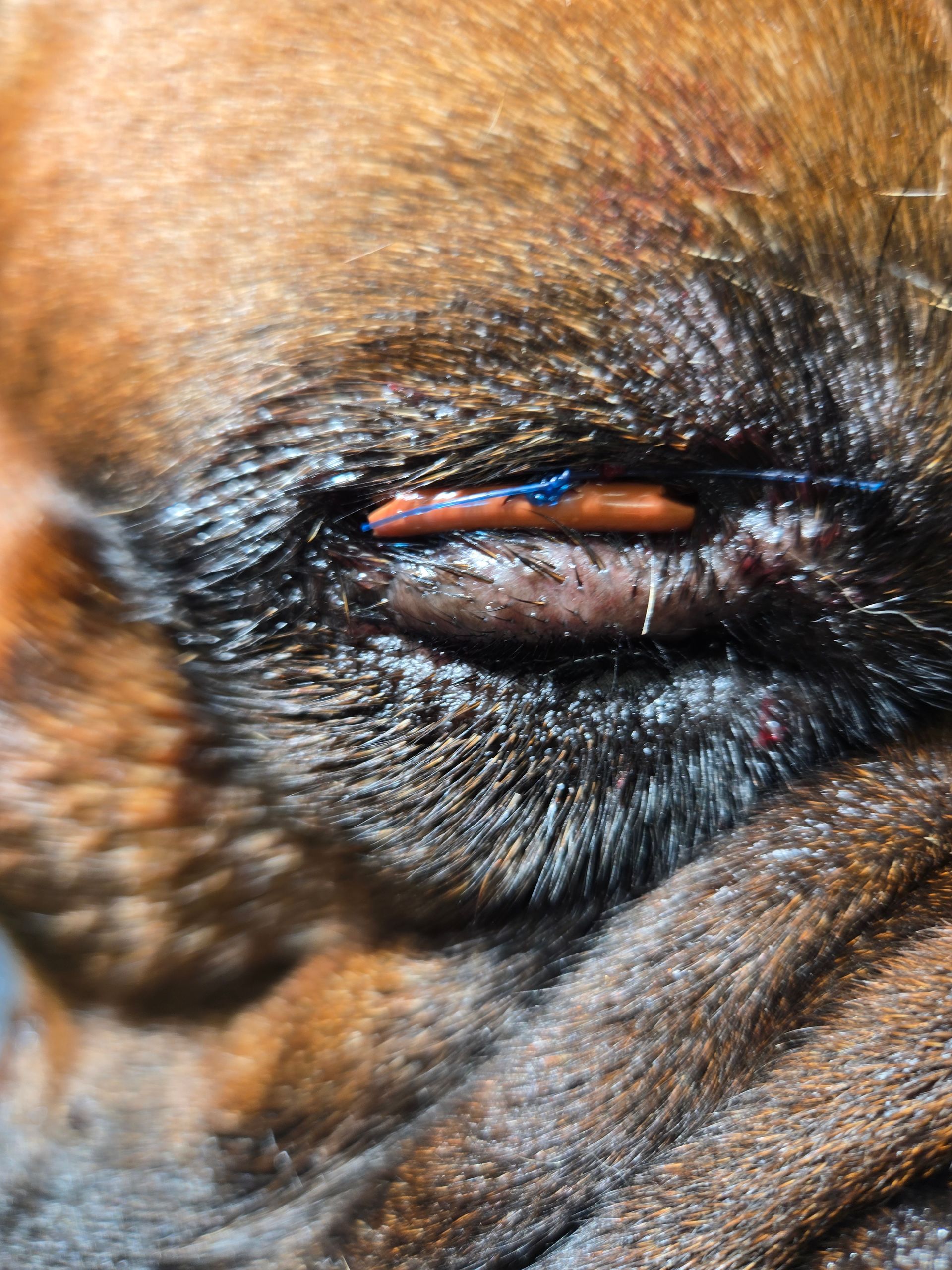
• Temporary protective eyelid closure, also known as a temporary tarsorrhaphy
• Placement of protective sutures or support to reduce exposure
• Treatment of traumatic wounds involving the tissues surrounding the eye
• Surgical management of selected globe-threatening injuries
A temporary tarsorrhaphy partially closes and supports the eyelids over the eye. It may be used to protect the cornea, reduce exposure, maintain globe position, and allow injured tissues to heal.
These procedures require careful judgment. The goal is not simply to improve the appearance of the eye. The goal is to relieve pain, protect viable tissues, reduce additional injury, and preserve the eye whenever that is medically realistic.
Not every severely injured eye can be saved, and preservation of the globe does not always mean that vision will return. Our team discusses the examination findings, treatment options, expected recovery, vision potential, comfort, and prognosis with the pet’s family before proceeding whenever circumstances permit.
RECHECKS ARE PART OF THE TREATMENT
Emergency ocular treatment does not end when the first procedure is completed.
Close follow-up is essential because swelling, intraocular pressure, corneal health, tissue viability, comfort, and vision can change during recovery.
Recheck examinations allow our team to:
• Monitor corneal healing
• Reassess pain and inflammation
• Evaluate ocular pressure when appropriate
• Check sutures and protective closures
• Determine whether the globe remains stable
• Adjust medications
• Decide when protective sutures may be removed
• Identify complications before they become more serious
• Coordinate ophthalmology referral when specialized care is needed
Some of the most meaningful ocular cases progress through several stages: emergency presentation, stabilization, imaging, surgical protection, and repeated follow-up until healing is established.
ADVANCED CARE UNDER ONE ROOF
Serious ocular injuries do not always occur in isolation. A patient may also require trauma evaluation, laboratory testing, advanced imaging, anesthesia, surgery, oxygen support, or hospitalization.
Pet ER is equipped to connect those services into one coordinated plan.
Our emergency ocular capabilities may include:
Urgent ocular evaluation
Pain control and medical stabilization
Fluorescein staining and detailed surface examination
Intraocular pressure assessment when appropriate
Ocular ultrasound
Eyelid, periocular, and protective ocular procedures when indicated
ICU monitoring and postoperative support
Serial rechecks and recovery monitoring
Coordination with veterinary ophthalmology when specialized care is required
This integrated approach allows us to evaluate the patient as a whole while giving the injured eye the immediate attention it requires.
WHAT TO DO BEFORE ARRIVING
If your pet has a protruding, swollen, bleeding, cloudy, or painful eye:
• Do not press on or attempt to reposition the eye yourself.
• Prevent your pet from rubbing or pawing at the face.
• Use an Elizabethan collar if one is immediately available and can be placed without causing further injury.
• Do not apply human eye drops, ointments, or leftover medications unless directed by a veterinarian.
• Call an emergency veterinary hospital immediately.
With ocular trauma, hours matter.
THE TAKE-HOME MESSAGE
A painful, red, swollen, protruding, cloudy, or suddenly injured eye deserves immediate veterinary attention.
Prompt evaluation may improve comfort, preserve treatment options, and increase the possibility of protecting the eye. When direct visualization is limited, ocular ultrasound can help us look deeper and make more informed decisions.
At Pet ER, emergency eye cases receive rapid assessment, advanced imaging when indicated, medical stabilization, selected ocular procedures, and close follow-up throughout recovery.
For emergency eye care or ocular ultrasound for dogs and cats, call Pet ER Small Animal Emergency Hospital at (631) 247-3837.
Please call ahead to confirm current doctor availability and emergency intake.